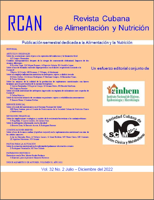
Sobre las asociaciones entre el peso a la captación del embarazo y la biometría fetal
Palabras clave:
Crecimiento fetal, Biometría fetal, Edad gestacional, Peso a la captación del embarazoResumen
Introducción: Un peso insuficiente en el momento de la captación del embarazo se puede trasladar a tasas menores del crecimiento fetal. Objetivo: Examinar las asociaciones entre el peso a la captación del embarazo e indicadores selectos del crecimiento fetal. Locación del estudio: Hospital Gineco-Obstétrico “Eusebio Hernández Pérez” (Marianao, La Habana, Cuba). Diseño del estudio: Transversal, analítico. Serie de estudio: Cien mujeres (Edad promedio: 25.5 ± 6.9 años; Edad gestacional (EG) promedio: 25.9 ± 3.9 semanas; EG < 28 semanas: 70 % vs. EG entre 28 – 36 semanas: 30 %) que ingresaron consecutivamente en el hospital entre los meses de Noviembre del 2019 y Agosto del 2020 (ambos inclusive). Métodos: Se examinaron las asociaciones entre el peso a la captación del embarazo, por un lado; y los indicadores del crecimiento, fetal por el otro. Los indicadores de crecimiento fetal se obtuvieron en cada trimestre del embarazo mediante ultrasonografía. Resultados: De acuerdo con el peso a la captación del embarazo, las mujeres fueron clasificadas como: Peso insuficiente: 19 %; Peso suficiente: 41 %; Sobrepeso: 24 %; Obesidad: 16 %; respectivamente. Los valores de los indicadores de la biometría fetal comprendidos entre los percentiles 10 – 90 de las tablas de referencias fueron como sigue: Diámetro biparietal (DBP): 49.5 %; Circunferencia cefálica (CC): 46.0 %; Circunferencia abdominal (CA): 59.3 %; Longitud del fémur (LF): 69.9 %; Peso fetal (PF): 44.2 %. Los indicadores del crecimiento fetal se comportaron de acuerdo con la edad gestacional como sigue: < 28 semanas: DBP: 54.6 ± 10.1 mm; CC: 204.7 ± 30.8 mm; CA: 181.0 ± 33.9 mm; LF: 40.1 ± 7.1 mm; PF: 587.1 ± 266.5 g; Entre 28 – 36 semanas: DBP: 72.5 ± 8.7 mm (D = +17.9 mm); CC: 259.2 ± 33.1 mm (D = +54.5 mm); CA: 249.0 ± 33.5 mm (D = +68.0 mm); LF: 56.5 ± 9.1 mm (D = +16.4 mm); PF: 1,648.9 ± 883.5 g (D = +1,061.8 g; p < 0.05); respectivamente. Por su parte, los valores de los indicadores del crecimiento fetal disminuyeron según el peso de la mujer a la captación del embarazo: DBP: Peso insuficiente: 65.9 ± 11.2 mm (D = +5.2 mm); Peso suficiente: 60.7 ± 12.6 mm (D = 0.0 mm); Sobrepeso: 59.1 ± 12.3 mm (D = -1.6 mm); Obesidad: 55.9 ± 13.8 mm (D = -4.8 mm; p < 0.05); CC: Peso insuficiente: 239.7 ± 32.9 mm (D = +14.5 mm); Peso suficiente: 225.2 ± 40.0 mm (D = 0.0 mm); Sobrepeso: 215.7 ± 38.8 mm (D = -9.5 mm); Obesidad: 208.5 ± 44.4 mm (D = -16.7 mm; p < 0.05); CA: Peso insuficiente: 214.5 ± 39.1 mm (D = +8.1 mm); Peso suficiente: 206.4 ± 45.9 mm (D = 0.0 mm); Sobrepeso: 198.1 ± 44.8 mm (D = -8.3 mm); Obesidad: 190.3 ± 52.2 mm (D = -16.1 mm; p > 0.05); LF: Peso insuficiente: 48.3 ± 8.6 mm (D = +1.6 mm); Peso suficiente: 46.7 ± 11.1 mm (D = 0.0 mm); Sobrepeso: 53.6 ± 50.3 mm (D = +6.9 mm); Obesidad: 41.7 ± 11.4 mm (D = -5.0 mm; p < 0.05); Peso fetal: Peso insuficiente: 985.4 ± 473.9 g (D = +89.8 g); Peso suficiente: 895.6 ± 504.1 g (D = 0.0 g); Sobrepeso: 841.4 ± 588.3 g (D = -54.2 g); Obesidad: 745.4 ± 625.7 g (D = -150.2 g; p > 0.05); respectivamente. El cambio observado en el indicador fetal fue solo explicado por la edad gestacional de la embarazada. La influencia sobre la biometría fetal del peso de la mujer en la captación del embarazo fue (cuando más) marginal. Conclusiones: En el momento actual, el comportamiento de los indicadores del crecimiento fetal es solo dependiente de la edad gestacional.Citas
Géa-Horta T, Silva Rde C, Fiaccone RL, Barreto ML, Velásquez-Meléndez G. Factors associated with nutritional outcomes in the mother-child dyad: A population-based cross-sectional study. Public Health Nutr 2016;19(15):2725-33. Disponible en: http://doi:10.1017/S136898001600080X. Fecha de última visita: 17 de Febrero del 2022.
Monterrosa EC, Pelto GH. The mother-child food relationship in the study of infant and young child feeding practices. Sight Life 2017;31:46-51.
Marshall NE, Abrams B, Barbour LA, Catalano P, Christian P, Friedman JE; et al. The importance of nutrition in pregnancy and lactation: Lifelong consequences. Am J Obstet Gynecol 2022;226:607-32.
Shapira N. Prenatal nutrition: A critical window of opportunity for mother and child. Women’s Health 2008;4:639-56.
Alabduljabbar S, Zaidan SA, Lakshmanan AP, Terranegra A. Personalized nutrition approach in pregnancy and early life to tackle childhood and adult non-communicable diseases. Life [Basel] 2021;11(6):467. Disponible en: http://doi:10.3390/life11060467. Fecha de última visita: 17 de Febrero del 2022.
Calcaterra V, Cena H, Verduci E, Bosetti A, Pelizzo G, Zuccotti GV. Nutritional surveillance for the best start in life, promoting health for neonates, infants and children. Nutrients 2020;12(11):3386. Disponible en: http://doi:10.3390/nu12113386. Fecha de última visita: 17 de Febrero del 2022.
Baird J, Jacob C, Barker M, Fall CH, Hanson M, Harvey NC; et al. Developmental origins of health and disease: A lifecourse approach to the prevention of non-communicable diseases. Healthcare [Basel] 2017;5(1):14. Disponible en: http://doi:10.3390/healthcare5010014. Fecha de última visita: 18 de Febrero del 2022.
Hanson M, Godfrey KM, Lillycrop KA, Burdge GC, Gluckman PD. Developmental plasticity and developmental origins of non-communicable disease: theoretical considerations and epigenetic mechanisms. Prog Biophys Mol Biol 2011;106(1):272-80. Disponible en: http://doi:10.1016/j.pbiomolbio.2010.12.008. Fecha de última visita: 18 de Febrero del 2022.
Bianchi ME, Restrepo JM. Low birthweight as a risk factor for non-communicable diseases in adults. Front Med [Lausanne] 2022;8:793990. Disponible en: http://doi:10.3389/fmed.2021.793990. Fecha de última visita: 18 de Febrero del 2022.
Zanardo V, Fanelli T, Weiner G, Fanos V, Zaninotto M, Visentin S; et al. Intrauterine growth restriction is associated with persistent aortic wall thickening and glomerular proteinuria during infancy. Kidney Int 2011;80:119-23.
Albu AR, Anca AF, Horhoianu VV, Horhoianu IA. Predictive factors for intrauterine growth restriction. J Med Life 2014;7:165-71.
Benítez Martín A, Vargas Pérez M, Manzanares Galán S. Ultrasound and biochemical first trimester markers as predictive factors for intrauterine growth restriction. Obstet Gynaecol Cases 2017;4:6-10.
Haggag A, Hamisa M, Elsayed-Atallah W, Marie S. Accuracy of transcerebellar diameter in comparison with biparietal diameter, femur length, and fetal kidney length in the sonographic assessment of gestational age in the third trimester of pregnancy. Tanta Medical J 2022;50(3):199. Disponible en: https://www.tdj.eg.net/article.asp?issn=1110-1415;year=2022;volume=50;issue=3;spage=199;epage=203;aulast=Haggag. Fecha de última visita: 18 de Febrero del 2022.
Agbaje MA, Alao AI, Owonikoko KM. Ultrasonographic foetal head circumference and cheek-to-cheek diameter at term as predictors of labour outcomes. Nigerian Postgrad Med J 2022;29:123-30.
Li J, Xu H, Shen M, Li S, Wang L, Lu Y, Li Q. Etiologies and adverse outcomes of fetuses with short femur length based on proportion and percentile categorization. Adv Ultrasound Diagn Ther 2022;6:7-13.
Andrade KAP. Abdominal circumference cut-off point: An overview. Arch Venezolanos Farmacol Ter 2022;41:299-306.
Aye AA, Agida TE, Babalola AA, Isah AY, Adewole ND. Accuracy of ultrasound estimation of fetal weight at term: A comparison of Shepard and Hadlock methods. Ann Afr Med 2022;21 (1):49-53. Disponible en: http://doi:10.4103/aam.aam_76_20. Fecha de última visita: 18 de Febrero del 2022.
Dudley NJ. A systemic review of the ultrasound estimation of foetal weight. Ultrasound Obstet Gynecol 2005;25:80-9.
Albouy‐Llaty M, Thiebaugeorges O, Goua V, Magnin G, Schweitzer M, Forhan A; et al.; for the EDEN Mother-Child Cohort Study Group. Influence of fetal and parental factors on intrauterine growth measurements: Results of the EDEN mother-child cohort. Ultrasound Obstet Gynecol 2011;38:673-80.
Gómez Mendoza C, Ruiz Álvarez P, Garrido Bosze I, Rodríguez Calvo MD. Bajo peso al nacer, una problemática actual. AMC 2018;22(4):408-16. Disponible en: http://scielo.sld.cu/scielo.php?script=sci_arttext&pid=S1025-02552018000400408&lng=es. Fecha de última visita: 18 de Febrero del 2022.
López González A. Sobre los factores de riesgo del bajo peso al nacer. RCAN Rev Cubana Aliment Nutr 2020;30:195-217.
González AL, Suárez AR, Cámbara AC, Gómez RF. Eventos maternos asociados al bajo peso al nacer en un municipio de la ciudad de La Habana. RCAN Rev Cubana Aliment Nutr 2019;29:64-84.
Monagas Travieso DM. Bajo peso al nacer y salud materna. La experiencia de un policlínico universitario. RCAN Rev Cubana Aliment Nutr 2021;31:425-38.
Rubio NR, Batancour EM, Rodríguez VIF, Rubio NR, Raymond YV, Socorro JJ. Identificación de competencias específicas de enfermería para el cuidado del recién nacido en recuperación nutricional, Hospital “Eusebio Hernández Pérez”. 2020. Rev Uruguaya Enfermería 2022;17(1):1-13. Disponible en: http://rue.fenf.edu.uy/index.php/rue/article/view/343. Fecha de última visita: 19 de Febrero del 2022.
Díaz ME, Montero M, Jiménez S, Wong I, Moreno V. Tablas antropométricas para la evaluación de la mujer embarazada. Boletín número 3. Consejo Nacional de Sociedades Científicas de la Salud. La Habana: 2009. Disponible en: http://files.sld.cu/boletincnscs/files/2009/11/respub2009dramaria-elena.pdf. Fecha de última visita: 19 de Febrero del 2022.
Colectivo de autores. Obstetricia y perinatología. Diagnóstico y tratamiento. Editorial Ciencias Médicas. La Habana: 2012.
Santana Porbén S, Canalejo Martínez H. Manual de Procedimientos Bioestadísticos. Editorial EAE Académica Española. Madrid: 2012.
Draper NR, Smith H. Applied regression analysis. Volumen 326. John Wiley & Sons. New York: 1998.
Dudink I, Hüppi PS, Sizonenko SV, Castillo-Melendez M, Sutherland AE, Allison BJ, Miller SL. Altered trajectory of neurodevelopment associated with fetal growth restriction. Exp Neurol 2022;347:113885. Disponible en: http://doi:10.1016/j.expneurol.2021.113885. Fecha de última visita: 19 de Febrero del 2022.
Langley‐Evans SC, Pearce J, Ellis S. Overweight, obesity and excessive weight gain in pregnancy as risk factors for adverse pregnancy outcomes: A narrative review. J Human Nutr Diet 2022;35:250-64.
Fakhraei R, Denize K, Simon A, Sharif A, Zhu-Pawlowsky J, Dingwall-Harvey ALJ; et al. Predictors of adverse pregnancy outcomes in pregnant women living with obesity: A systematic review. Int J Environ Res Public Health 2022;19(4):2063. Disponible en: http://doi:10.3390/ijerph19042063. Fecha de última visita: 20 de Febrero del 2022.
Cnattingius S, Villamor E, Lagerros YT, Wikström AK, Granath F. High birth weight and obesity- A vicious circle across generations. Int J Obes 2012;36;1320-4.
Gaudet L, Ferraro ZM, Wen SW, Walker M. Maternal obesity and occurrence of fetal macrosomia: A systematic review and meta-analysis. Biomed Res Int 2014;2014:640291. Disponible en: http://doi:10.1155/2014/640291. Fecha de última visita: 20 de Febrero del 2022.
McDonald SD, Han Z, Mulla S, Beyene J; for the Knowledge Synthesis Group. Overweight and obesity in mothers and risk of preterm birth and low birth weight infants: Systematic review and meta-analyses. BMJ 2010;341:c3428. Disponible en: http://doi:10.1136/bmj.c3428. Fecha de última visita: 20 de Febrero del 2022.
Bonnet D. Impacts of prenatal diagnosis of congenital heart diseases on outcomes. Transl Pediatr 2021;10(8):2241-9. Disponible en: http://doi:10.21037/tp-20-267. Fecha de última visita: 20 de Febrero del 2022.
Kalinderi K, Delkos D, Kalinderis M, Athanasiadis A, Kalogiannidis I. Urinary tract infection during pregnancy: Current concepts on a common multifaceted problem. J Obstet Gynaecol 2018;38:448-53.
Naik VD, Lee J, Wu G, Washburn S, Ramadoss J. Effects of nutrition and gestational alcohol consumption on fetal growth and development. Nutr Rev 2022;80:1568-79.
Tarasi B, Cornuz J, Clair C, Baud D. Cigarette smoking during pregnancy and adverse perinatal outcomes: A cross-sectional study over 10 years. BMC Public Health 2022;22(1):2403. Disponible en: http://doi:10.1186/s12889-022-14881-4. Fecha de última visita: 20 de Febrero del 2022.


